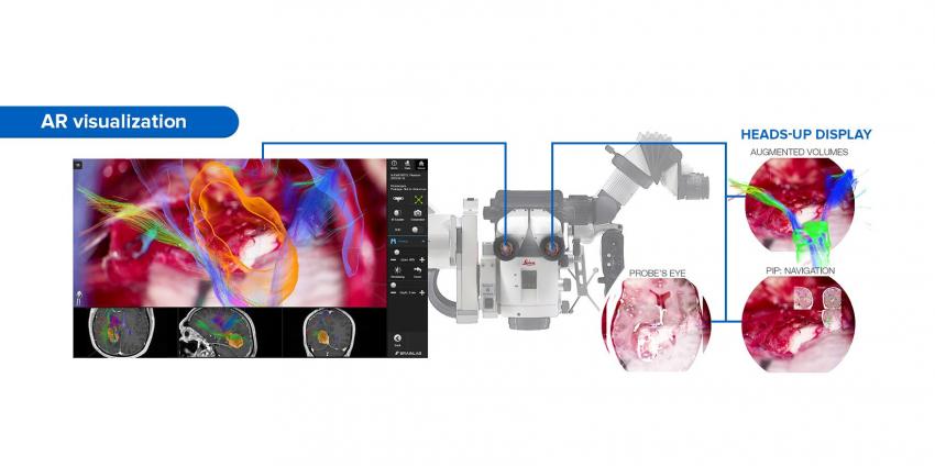
Leica introduces new AR visualization technologies for surgical microscopes
Neurosurgery is developing rapidly and enabling doctors to solve health problems that might seem impossible to solve at first. Modern microscopes allow obtaining the most accurate information, but sometimes it is not enough, as a few seconds delay in data processing can cost a human life. That's exactly why Leica decided to design a completely new image processing technology — AR (augmented reality). This is a significant step forward as it allows diagnosing patients and making treatment decisions quickly and easily.

Microscopic systems by Leica Microsystems bring medicine to a completely new level, being the first in the world to receive FDA 510(k) clearance. Due to modern products, doctors can conduct intraoperative angiography and use three types of fluorescence at the same time. One of such products is Leica GLOW800 system, which enhances the microscope view due to AR visualization technology.
Technical characteristics of the device
The new surgical microscope was first introduced at AANS (American Association of Neurological Surgeons) meeting in April 2017. Since then, the popularity of the new product has been growing and the whole world has started taking interest in it.
The new product has a number of specifications and new AR image processing technologies are among them. They ensure high-quality visualization, which will enable doctors to make right decisions when working with blood vessels, including arteriovenous malformations and aneurysms.
Leica GLOW800 system is a new method of vascular fluorescence, which is used in neurosurgery. It has a range of specifications:
- CaptiView HD image injection system;
- 3D visualization;
- possibility to display three types of fluorescence.

The president of Leica, Markus Lusser, states that the functional capacity of a microscope is not enough for neurosurgery, which is why it is necessary to develop the most powerful equipment possible. This was exactly what pushed the company to develop the new GLOW800 system which can be connected to any microscope in any hospital.
The advantages of AR visualization
Visualization with the help of GLOW800 combines the contrast of fluorescent imaging with the full visual spectrum of white light. As a result, the surgeon will obtain the white light image with the colored fluorescence flow in real-time. AR technology allows the surgeon to see and have full account of the structure through the microscope without switching eyepieces or orientation.
In addition, CaptiView HD microscope visualization allows obtaining data in high resolution directly through the microscope with no need to look at the screen. It allows the surgeon to work without interruption, while having accurate information about the condition of the patient’s organ, which is necessary to make the right decision regarding treatment.

The device was tested in combination with the popular M530 microscope. Integration of functional capabilities of these two systems allows visualization of brain anatomy in real-time with high contrast and white light. This allows the surgeon to diagnose with the highest accuracy during vascular neurosurgery, and creates an opportunity to activate different modes of fluorescence with the help of a microscope handle or a special foot pedal, maintaining visual contact with the patient’s organ at the same time.
BiMedis Сompany
29.10.2017