New PET/CT imaging method improves cardiac sarcoidosis diagnostics
As of today, the quality diagnostics of rare diseases remains an important issue, and cardiac sarcoidosis is one of such conditions.
Sarcoidosis is an inflammatory disease characterized by the appearance of epithelioid granulomas which commonly affect several organs, in particular:
- heart;
- lungs;
- liver;
- lymph nodes, etc.
A new diagnostic technique, which helps to reveal cardiac sarcoidosis and provide more accurate data than conventional tests, was presented to the Journal of Nuclear Cardiology readers by researchers from the University of Illinois, Chicago.

Revolutionary PET/CT scanning technology
Examinations, conducted employing positron emission tomography (PET) or computed tomography, were not always understandable enough. It is usually difficult to interpret the combination of data, received even from both devices, which may eventually lead to erroneous conclusions. That is why researchers from the UIC Medical College developed a new technology, combining positron emission and computed tomography (PET/CT). This technique allowed doctors to obtain better and crisper images, facilitating more accurate cardiac sarcoidosis detection in those patients who kept to an ordinary 24-hour diet. A 72-hour diet with increased amounts of fat and a minimum level of sugar was commonly necessary to conduct such kind of examinations. The scientists used this protocol to study the cardiac sarcoidosis relation to other organs sarcoidosis development.
New PET/CT protocols investigation results

The studies were conducted on 188 patients during a year (from December 2014 to December 2015). The entire investigated group followed the aforementioned diet for 72 hours before the diagnostic procedures and underwent a complete PET/CT scan at a hospital in the University of Illinois.
The study results showed that the new PET/CT scan protocol provides an accurate diagnosis, revealing not only cardiac sarcoidosis. The researchers claim that eight out of the twenty patients diagnosed with cardiac sarcoidosis were also diagnosed with sarcoidosis in other organs.
Dr. Nadera Sweiss, a professor of rheumatology at the UIC Medical College and one of the investigation authors said, “Knowing there is disease in organs other than the heart changes the way we approach treatment — it allows us to more accurately stage the disease and treat it accordingly”.

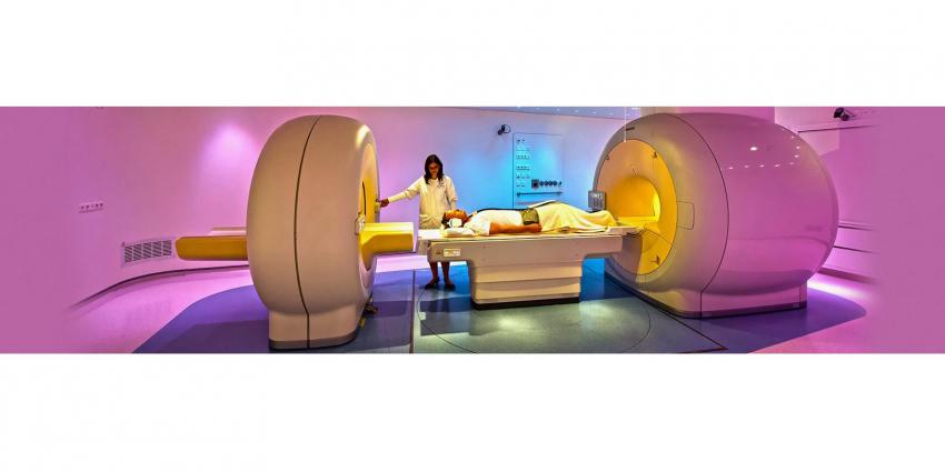
PET/CT diagnostics is an innovative technique that combines positron emission tomography, capable of visualizing the human body’s functional abnormalities and computed tomography. Using this kind of diagnostics, we receive detailed information about the human body’s anatomical structure. Due to the precise and quality data, which is possible to obtain using a PET/CT scanner, this type of examination is increasingly utilized to diagnose various diseases.
On the BiMedis ads platform, you can find out more about a wide range of radiological equipment, such as PET and CT scanners, etc. The right choice of powerful equipment is the key factor to quality diagnostics and timely treatment.
21.09.2017