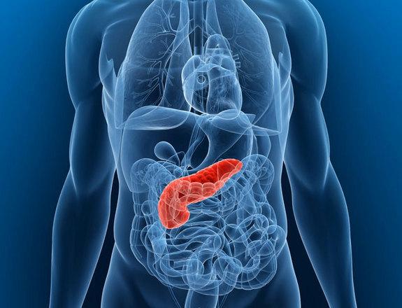
RESEARCHERS USE MRI TO VISUALIZE CHANGES IN PANCREAS IN EARLY STAGE DIABETES
Scientists from Joslin Diabetes Center have published a novel study concerning the origin of type I diabetes and the ability of magnetic resonance imaging (MRI) to help visualize inflammation in the pancreas. Knowledge about pancreatic inflammation and its consequences may help diagnose and treat type I diabetes in a timely manner, and may shed some light about disease progression.
The study was performed using ferumoxytol, a coated iron nanoparticle, used in iron replacement therapy and MRI. As previously published in similar studies, ferumoxytol leaks out of blood vessels in areas of inflammation and is subsequently taken up by immune cells that are responsible for phagocytosis, macrophages, which converge at site of inflammation.
The underlying factors for the development of type I diabetes are autoimmune reactions and inflammation of the pancreas. Although tests for autoantibodies (antibodies directed against the pancreas) can be an indicator of immune response, they are not specific enough to show the chance of developing diabetes.
"Many people have genetic variants that put them at risk for type 1 diabetes," explains study colead author Jason Gaglia, M.D., M.M.Sc., an Assistant Investigator in the Section of Immunobiololgy at Joslin. "Some develop autoimmunity, but only a small number develop clinical disease."
The study included 11 patients with recently diagnosed type I diabetes and positive for autoantibodies, and 10 control patients with no signs or family history of diabetes.
The MRI techniques used to study the pancreas were adapted from MRI mapping algorithms originally developed for whole brain scanning. Gaglia noted that the ferumoxytol dose used in the study was approximately one quarter of the recommended therapeutic dose for iron replacement therapy. So, other than the unique MRI mapping algorithms, the materials used in the study are widely available and can be repeated in any center.
The potential applications for this imaging technique could help aid diabetes research to show a complete picture of the development of type I diabetes. Gaglia says, the ferumoxytol MRI images revealed that in their patient group, "inflammation was not uniform across the entire pancreas. There was also a large amount of variation between individuals, which aligns with what you see clinically. That’s never been shown in living humans before."
Seeing as how not everybody with autoantibodies develops diabetes, using this diagnostic approach, physicians can better identify patients who are more likely to progress to diabetes, and classify them according to treatment strategies.
02.04.2015